Diagnosis and Treatments of Retinal Detachment
Retinal detachment is a serious eye condition that occurs when the retina—the light-sensitive layer of tissue at the back of the eye—pulls away from its normal position. If left untreated, retinal detachment can lead to permanent vision loss. Early diagnosis and prompt treatment are critical to preserving vision. In this article, we’ll explore how retinal detachment is diagnosed and the treatment options available.
What is Retinal Detachment?
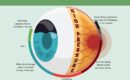
Retinal detachment occurs when the retina separates from the underlying layers of the eye that supply it with oxygen and nutrients. This separation disrupts the retina’s ability to function, leading to vision loss. There are three main types of retinal detachment:
- Rhegmatogenous Retinal Detachment: The most common type, caused by a tear or hole in the retina that allows fluid to seep underneath.
- Tractional Retinal Detachment: Occurs when scar tissue on the retina’s surface pulls it away from the back of the eye.
- Exudative Retinal Detachment: Caused by fluid buildup beneath the retina due to inflammation, injury, or other underlying conditions.
Symptoms of Retinal Detachment
If you experience any of the following symptoms, seek immediate medical attention:
- Sudden appearance of floaters (dark spots or squiggly lines in your vision).
- Flashes of light in one or both eyes.
- A shadow or curtain-like effect in your peripheral or central vision.
- Blurred or distorted vision.
Diagnosis of Retinal Detachment
Early diagnosis is crucial for successful treatment. Here’s how retinal detachment is diagnosed:
- Comprehensive Eye Exam: Your eye doctor will examine your retina using an ophthalmoscope, a tool that allows them to see the back of your eye.
- Ultrasound Imaging: If the retina cannot be seen clearly due to bleeding or other obstructions, an ultrasound may be used to create images of the retina.
- Optical Coherence Tomography (OCT): This non-invasive imaging test provides detailed cross-sectional images of the retina, helping to identify the extent of detachment.
- Fluorescein Angiography: A dye is injected into the bloodstream, and images are taken to assess blood flow in the retina and identify any abnormalities.
Treatment Options for Retinal Detachment
The treatment for retinal detachment depends on the type and severity of the condition. The goal is to reattach the retina and restore vision. Here are the most common treatment options:
- Laser Surgery (Photocoagulation): Used for small retinal tears or holes. A laser is used to create small burns around the tear, forming scar tissue that seals the retina to the underlying tissue.
- Cryopexy (Freezing Treatment): A freezing probe is applied to the outer surface of the eye to create scar tissue around the retinal tear, sealing it in place.
- Pneumatic Retinopexy: A gas bubble is injected into the vitreous cavity of the eye. The bubble pushes the retina back into place, and laser or cryopexy is used to seal the tear. Patients must maintain a specific head position for several days to keep the bubble in place.
- Scleral Buckling: A silicone band is placed around the eye to gently push the wall of the eye against the detached retina, allowing it to reattach. This is often combined with cryopexy or laser treatment.
- Vitrectomy: A surgical procedure where the vitreous gel (the gel-like substance inside the eye) is removed and replaced with a gas or silicone oil bubble to push the retina back into place. The gas bubble eventually dissipates, but silicone oil may require a second surgery for removal.
Recovery and Prognosis
- The success of retinal detachment treatment depends on the extent of the detachment and how quickly it is treated.
- Vision recovery may take weeks to months, and some patients may not regain full vision, especially if the macula (central part of the retina) was affected.
- Follow-up appointments are essential to monitor healing and prevent complications.
When to See a Specialist
If you suspect retinal detachment, don’t wait—seek help immediately. At Sood Eye Care, we specialize in diagnosing and treating retinal conditions. Our team, led by Dr. Saurabh Sood, is equipped with the latest technology and expertise to provide the best possible care for your eyes.